
[vc_empty_space height=”20px”]
超高速レーザは、新しいタイプの非線形顕微鏡技術を可能にし、顕微鏡の分野に革命をもたらしました。 フェムト秒パルスレーザは走査型レーザ顕微鏡(LSM)で非線形光学プロセスを利用するために、ハイピークパワー(1-100 kW)と低平均出力(10-1000 mW)の理想的な組み合わせを提供します。
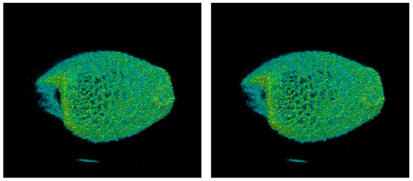
図1:コンバラリア花粉のステレオ2光子蛍光像
LSMの作業の大部分は、青色および緑色のレーザ光源を使用した通常の単一光子蛍光を使用して行われています。 しかし非線形顕微鏡法は、単一光子蛍光法に比べて確かなメリットがあります。
- 共焦点ピンホールを必要としない自然な断層画像
- 長波長の使用により散乱媒体へより深く侵入する
- 励起体積が小さいため光毒性が少なく、生きた細胞や生物の観察時間が長くなります。
このような利点が生物学・医学・神経科学の研究者の間でマルチフォトンLSMの人気が高まっている理由を説明しています。 ここでは非線形顕微鏡において2つの代表的な2光子顕微鏡と第3高調波(THG)イメージングについて説明します。 我々は様々なタイプのモードロックファイバレーザでこれらの技術を使用した画像を示します。
二光子顕微鏡 二光子顕微鏡法は、通常は使用した二光子の半分の波長で吸収している分子に、同じような波長の二光子を同時に吸収させる多光子プロセスに基づいています。そして分子は励起を失い、元の吸収された光子の周波数の和よりも低い周波数(より長い波長)で別の光子を放出します。
例えばローダミンのような色素は、緑色の波長(〜530 nm)で強い吸収を持ち、そしてより長い波長(〜600 nm)で蛍光を発します。 2光子蛍光を励起するには波長1060 nmのレーザパルスを使用することができます。 通常の一光子吸収プロセスによって1060nmの光の吸収は事実上ありませんが、1060nmでの2つの光子は、530nmの単一光子でローダミン分子を励起するのと本質的に同じ効果で同時に吸収することができます。 この結果として、分子の通常の発光バンド内で蛍光が発生します。 しかし蛍光量がポンプ強度に直線的に比例する1光子蛍光とは異なり、2光子蛍光量は式1に見られるようにポンプ強度の2乗に比例します。
F1-phoα k1-phoI 1.
F2-pho α k2-phoI2 1.b.
2光子吸収の係数は非常に小さく、プロセスを駆動するためには高いピークパワーと非常に小さな焦点が必要になります。 またビームの焦点位置のウエスト部分で非常に局所的に励起され、それが固有の断層画像を与えています。
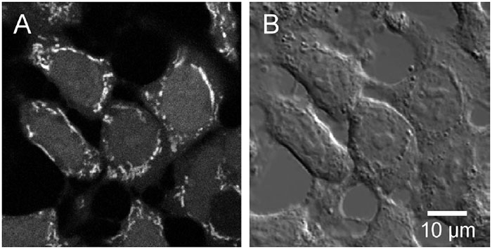
図2:A)インド-1 AMを装填したHT¬29細胞の2光子励起蛍光像。B)2光子励起光と同時に記録された示差干渉コントラスト画像(提供:Roland Nitschke、フライブルク大学
TiSapphireレーザは波長可変性(700-950 nm)と高い平均出力(~1ワット)により、二光子顕微鏡用に最もポピュラーな光源です。 しかし波長可変性や高出力を必要としないアプリケーションも数多く存在します。 実際には試料に照射する光パワーの量の目標値は10mW以下であるべきです。高いパワーはサンプルに損傷を与える可能性があり、またはその寿命を短縮させる可能性があります。 高出力なレーザは顕微鏡システムにおける光学損失を補償するために使用されます。顕微鏡の光学系は通常は近赤外励起波長ではなく可視波長に最適化されているため、近赤外励起波長において光学損失が大きいからです。ます。 さらに単一の波長であっても、様々な機能的なイメージングオプションを提供する多数の蛍光標識試薬の蛍光色素が利用可能です。
図2は、1998年にIMRA Femtolite A-15フェムト秒ファイバーレーザを使用して撮影したHT29(ヒト結腸癌)細胞の微分干渉観察(DIC)と2光子蛍光画像です。1 細胞はカルシウムイオン濃度と活性を明らかにするためにIndo-1で染色しました。 他の生物の画像もそれらの自然な自家蛍光を介して染色せずに撮れました。 図1の立体画像は同じ二光子LSMシステムで撮影した40の断層画像から構築されたもので、またFemtolite A-15を搭載しています。 最初に市販されたフェムト秒ファイバーレーザの1つであるFemtolite A-15は780nmで平均出力15mW、パルス持続時間200fsでした。 最近のIMRA製品は堅牢でコンパクトなパッケージで性能が大幅に向上しています。 例えばFemtolite FX-100iは、わずか14 x 10 cm2のフットプリントサイズで、805 nmで100 mW以上の平均出力を提供します。
第三高調波(THG)イメージング
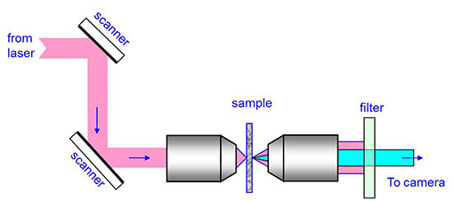
図3:THG顕微鏡システムの概略図。サンプルは透過型ジオメトリにセットされています。サンプル後の光学フィルターはポンプ光を除去し、THG信号光を通過させます。
多光子顕微鏡法におけるエキサイティングな新展開として、第三高調波発生(THG)顕微鏡法の実証があります2。 この手法では試料に入射したレーザパルスが第三高調波を発生させます。 このプロセスは対称性の制約により、通常、等方性のバルクでは起こりません。しかし異なる媒体間の表面では対称性が破られ、高調波が発生することがよく知られています。 このようにレーザからの強い光の下では、界面で最大の発光が起こり、これが最も興味深い特徴となります。 図3は試料にレーザを照射した場合の基本的な光学系を示しています。 THG光は透過方向に型で生成されるため、サンプルは、THG光を再度コリメート光にしてイメージングカメラに送るように配置設定されています。 二光子イメージングの場合と同様に、この技術では断層画像機能を提供します。 図4はFemtoliteモデルB-35から1560 nmでの励起パルスを使用して、第三高調波(THG)発生で撮影した葉の細胞の断面画像を示しています。 蛍光イメージングとは異なり、THGイメージングは色素での染色を必要としないので、光退色を排除することができ、研究者は一回当たり数時間、標本を観察することができます。
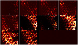
図4:ファイバーレーザーからの1560 nmのパルスを使用して撮影した葉の細胞の断面THG画像。提供:J. Squier、UCSD
もう一つの注目すべき波長領域は1000-1100 nmの領域です。 一般的な TiSapphire の 700~900 nm の波長領域と比較して、これらの長い波長は透過率が高く散乱も少ないため、さまざまな種類の組織をよりよく観察することができます。 IMRAのイッテルビウムをドープしたFCPA µJewel D-400フェムト秒ファイバーレーザは1041 nmで400 mWの平均出力を提供し、この種の顕微鏡に非常に適した光源となっています。
概要 モードロック型フェムト秒ファイバーレーザは二光子LSMや第3高調波発生(THG)顕微鏡など、様々なタイプの非線形顕微鏡に最適な光源です。 IMRAのエルビウムおよびイッテルビウムをドープしたフェムト秒ファイバーレーザは数種類の波長(780・805・1041・1560・1620 nm)で安定した高強度パルスを提供し、その小型で堅牢な形状ゆえに、これらのイメージング技術をほぼすべての環境で整備することができます。
参考文献
- Ricken, J. Leipziger, R. Gregor, R. Nitschke (1998)."HT29細胞における細胞質およびミトコンドリアCa2+過渡現象の同時測定" J. Biolog.Chem.第273巻第52号、pp. 34961-34969
- C.このように、このような研究では、研究者たちの研究室での研究の成果を、より多くの人に知ってもらうために、研究者たちの研究室での研究の成果を紹介しています。"このように、本研究では、このような研究成果をもとにして、研究者の皆様の研究活動を支援することを目的とした研究を行っています。38, No.26, pp.7393-7397
*IMRAは、Femtolite FX-100iまたはFemtolite Ultraファイバーレーザーの製造を終了しました。 類似製品については、当社のFemtolite F/G/Hラインをご覧ください。